miRNAs (microRNAs) participate in many diseases including cardiovascular disease. In contrast with our original hypothesis, miRNAs exist in circulating blood and are relatively stable due to binding with other materials. The aim of the present translational study is to establish a method of determining the absolute amount of an miRNA in blood and to determine the potential applications of circulating cell-free miR-1 (microRNA-1) in AMI (acute myocardial infarction). The results revealed that miR-1 is the most abundant miRNA in the heart and is also a heart- and muscle-specific miRNA. In a cardiac cell necrosis model induced by Triton X-100 in vitro, we found that cardiac miR-1 can be released into the culture medium and is stable at least for 24 h. In a rat model of AMI induced by coronary ligation, we found that serum miR-1 is quickly increased after AMI with a peak at 6 h, in which an increase in miR-1 of over 200-fold was demonstrated. The miR-1 level returned to basal levels at 3 days after AMI. Moreover, the serum miR-1 level in rats with AMI had a strong positive correlation with myocardial infarct size. To verify further the relationship between myocardial size and miR-1 level, an IP (ischaemic preconditioning) model was used. The results showed that IP significantly reduced circulating miR-1 levels and myocardial infract size induced by I/R (ischaemia/reperfusion) injury. Finally, the levels of circulating cell-free miR-1 were significantly increased in patients with AMI and had a positive correlation with serum CK-MB (creatine kinase-MB) levels. In conclusion, the results suggest that serum miR-1 could be a novel sensitive diagnostic biomarker for AMI.
Circulating cell-free miR-1 in rats with or without acute myocardial infarction (AMI)
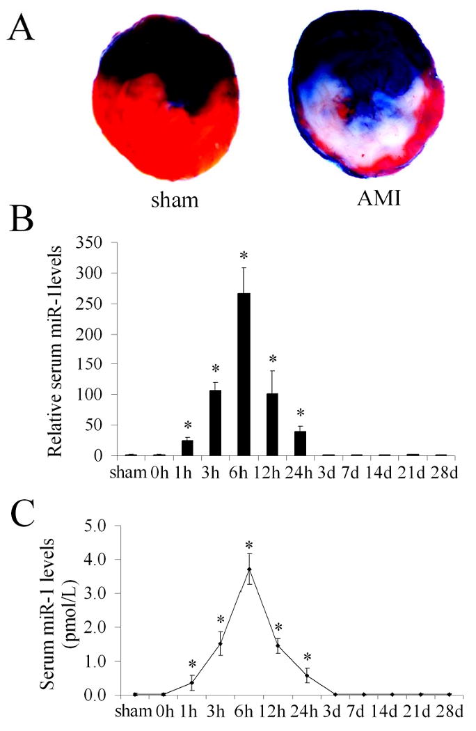
(A) Myocardial infarction in rat heart was induced by left anterior descending coronary artery ligation and the infarct size was determined by pathological staining in heart slices. Note: Color blue is Evans blue staining. The region without Evans blue staining is myocardial ischemic area at risk (IAR). Color red is the triphenyltetrazolium chloride (TTC) staining. TTC unstained area within IAR was the infarcted area. Infarct size is expressed as a percentage of the IAR (% IAR). (B) The relative serum miR-1 levels in 8 sham-opened control rats, and in 8 rats before (0 h) and at different time points after AMI. Note: the mean value of serum miR-1 before AMI was expressed as 1. n=8, *P<0.05 compared with the group before AMI (0 h). (C) The absolute amount of serum miR-1 sham-opened control rats, and in rats before (0 h) and at different time points after AMI. Note: n=8, *P<0.05 compared with the group before AMI.
Related Service
miRNA Microarray Service – LC Sciences provides a microRNA (miRNA) expression profiling service using microarrays based on our in-house developed µParaflo® technology platform. We have standard arrays for all mature miRNAs of all species available in the latest version of the miRBase database (Release 21, July 2014). Our service is comprehensive and includes sample labeling, array hybridization, image data processing and in-depth data analysis. Two-three weeks after receiving your total RNA samples, we’ll send you both the raw and fully analyzed data. [Learn more…]
Reference
Cheng Y, Tan N, Yang J, Liu X, Cao X, He P, Dong X, Qin S, Zhang C. (2010) A translational study of circulating cell-free microRNA-1 in acute myocardial infarction. Clinical Science 119, 87-95. [article]