Acute myocardial infarction (MI) due to coronary artery occlusion is accompanied by a pathological remodeling response that includes hypertrophic cardiac growth and fibrosis, which impair cardiac contractility.
Researchers at the University of Texas Southwestern Medical Center show by LC Sciences’ miRNA microarray that MI in mice and humans results in the dysregulation of specific miRNAs, which are similar to but distinct from those involved in hypertrophy and heart failure. Among the MI-regulated miRNAs are members of the miR-29 family, which are down-regulated in the region of the heart adjacent to the infarct.
Down-regulation of miR-29 with anti-miRs in vitro and in vivo induces the expression of collagens, whereas over-expression of miR-29 in fibroblasts reduces collagen expression. The researchers concluded that miR-29 acts as a regulator of cardiac fibrosis and represents a potential therapeutic target for tissue fibrosis in general.
MiRNA profiling in response to MI
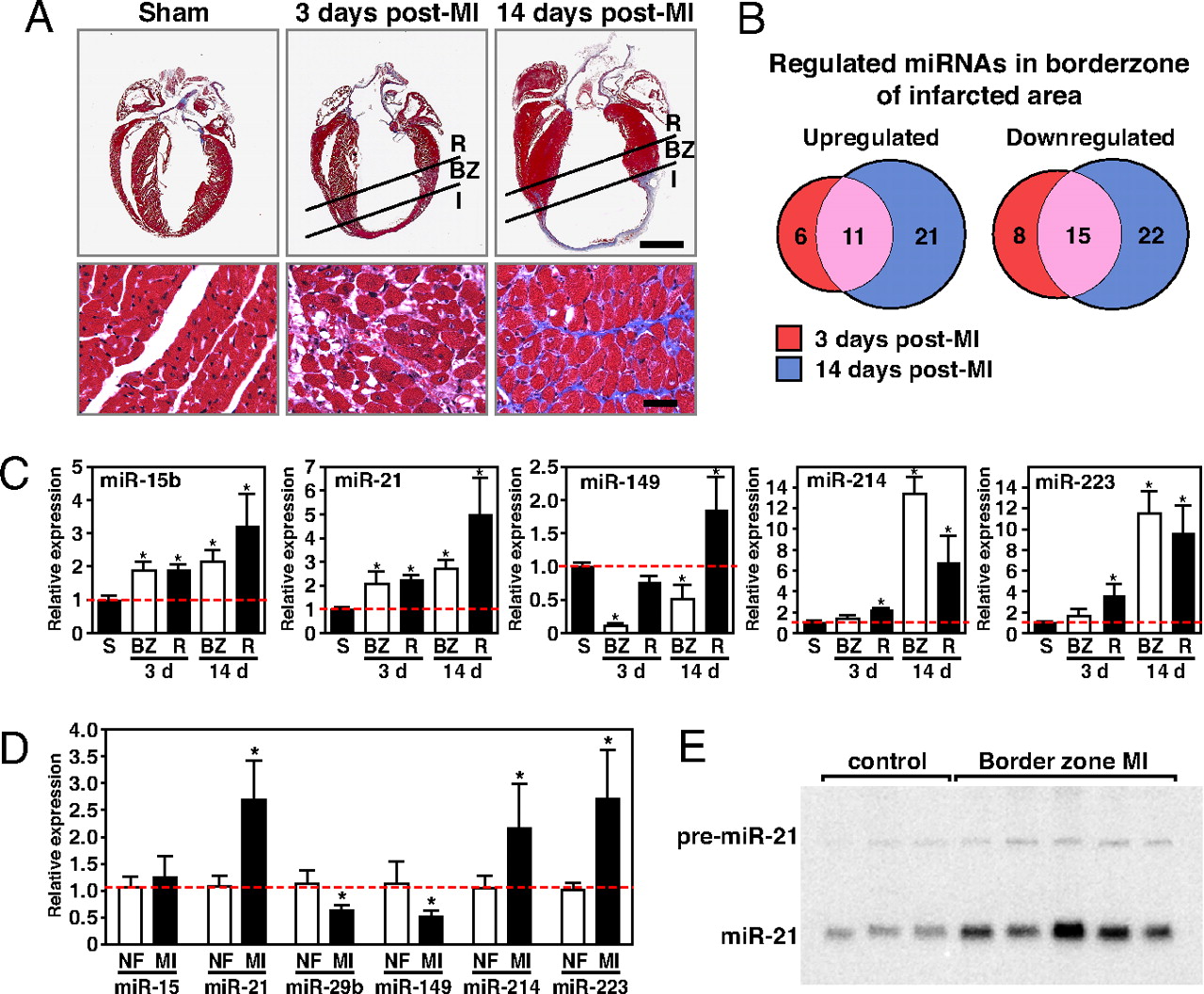
(A) Masson Trichrome staining of mouse heart sections shows early scar formation 3 days after MI, with myocyte hypertrophy, loss of myocytes, and collagen deposition. Fourteen days after MI, there is a thin stretched infarct that results in cardiac hypertrophy and interstitial fibrosis in the border zone of the infarcted region. I, infarct; BZ, borderzone; R, remote myocardium. (Lower) Higher magnification of the borderzone regions of the infarcted hearts and the comparable level of the sham operated heart. (Scale bars: Upper, 2 mm; Lower, 20 μm). (B) Microarray analysis reveals miRNAs are dynamically regulated in response to MI. Even after initial infarct healing, 14 days after MI, 11 miRs are overlappingly up-regulated, whereas 15 miRs are down-regulated. The number of miRNAs regulated ≥2-fold in each category is shown. (C) Real-time PCR analysis confirms the regulation of specific miRNAs in response to MI compared with sham operated animals (n = 3–4. S, sham; BZ, borderzone; R, remote. *, P < 0.05 compared with sham operated animals). (D) Real-time PCR analysis shows the regulation of miRNAs in human heart samples in response to MI compared with nonfailing hearts (n = 5–6, NF = nonfailing, MI = myocardial infarction). *, P < 0.05 compared with nonfailing hearts. (E) Northern blot analysis of three nonfailing human hearts and five human hearts after MI indicates a consistent increase in miR-21 in the borderzone of human heart samples in response to MI.
Related Service
miRNA Microarray Service – LC Sciences provides a microRNA (miRNA) expression profiling service using microarrays based on our in-house developed µParaflo® technology platform. We have standard arrays for all mature miRNAs of all species available in the latest version of the miRBase database (Release 21, July 2014). Our service is comprehensive and includes sample labeling, array hybridization, image data processing and in-depth data analysis. Two-three weeks after receiving your total RNA samples, we’ll send you both the raw and fully analyzed data. [Learn more…]
Reference
van Rooij E, Sutherland LB, Thatcher JE, DiMaio JM, Naseem RH, Marshall WS, Hill JA, Olson EN. (2008) Dysregulation of microRNAs after myocardial infarction reveals a role of miR-29 in cardiac fibrosis. Proc Natl Acad Sci USA 105(35), 13027-32. [article]