Several recent reports have suggested that microRNAs (miRNAs) might play critical roles in acute myocardial infarction (AMI). However, the miRNA expression signature in the early phase of AMI has not been identified. In this study, the miRNA expression signature was investigated in rat hearts at 6 h after AMI. Compared with the expression signature in the noninfarcted areas, 38 miRNAs were differentially expressed in infarcted areas and 33 miRNAs were aberrantly expressed in the border areas. Remarkably, miR-21 expression was significantly downregulated in infarcted areas, but was upregulated in border areas. The downregulation of miR-21 in the infarcted areas was inhibited by ischemic preconditioning, a known cardiac protective method. Overexpression of miR-21 via adenovirus expressing miR-21 (Ad-miR-21) decreased myocardial infarct size by 29% at 24 h and decreased the dimension of left ventricles at two weeks after AMI. Using both gain-of-function and loss-of-function approaches in cultured cardiac myocytes, we identified that miR-21 had a protective effect on ischemia-induced cell apoptosis that was associated with its target gene programmed cell death 4 (PDCD4) and activator protein 1 (AP-1) pathway. The protective effect of miR-21 against ischemia-induced cardiac myocyte damage was further confirmed in vivo by decreased cell apoptosis in the border and infarcted areas of the infarcted rat hearts after treatment with Ad-miR-21. The results suggest that miRNAs such as miR-21 may play critical roles in the early phase of AMI. Some miRNAs may be considered new therapeutic targets or even biomarkers for ischemic heart disease such as AMI.
miR-21 expression in different areas of the infarcted hearts and the effect of IP on the expression in the early phase of AMI.
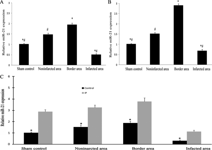
miRNAs were isolated in different areas of the infarcted hearts as well as in sham-opened hearts at 6 (A) and 24 h (B) after AMI. The expression of miR-21 was determined by qRT-PCR. miR-21 expression was down-regulated in the infarcted area at both 6 and 24 h after AMI compared with that in other areas and in sham-opened hearts. Notably, compared with that in other areas and sham-opened hearts, miR-21 expression in the border area was significantly increased. C, the effect of IP on miR-21 expression at 6 h after AMI. The IP was performed 6 h before the AMI. Note: Data presented as mean ± S.E. (error bars), n = 6, *, p < 0.05 compared with the noninfarcted control in A and C, and with the IP group in C. #, p < 0.05 compared with that in the border area.
Related Service
miRNA Microarray Service – LC Sciences provides a microRNA (miRNA) expression profiling service using microarrays based on our in-house developed µParaflo® technology platform. We have standard arrays for all mature miRNAs of all species available in the latest version of the miRBase database (Release 21, July 2014). Our service is comprehensive and includes sample labeling, array hybridization, image data processing and in-depth data analysis. Two-three weeks after receiving your total RNA samples, we’ll send you both the raw and fully analyzed data. [Learn more…]
Reference
Dong S, Cheng Y, Yang J, Li J, Liu X, Wang X, Wang D, Krall TJ, Delphin ES, Zhang C. (2009) MicroRNA expression signature and the role of microRNA-21 in the early phase of acute myocardial infarction. J Biol Chem 284(43):29514-25. [article]